Quick Facts
The microvilli of pigment cells interact with the tips of the rod and cone photoreceptor outer segments.
Structure and/or Key Features
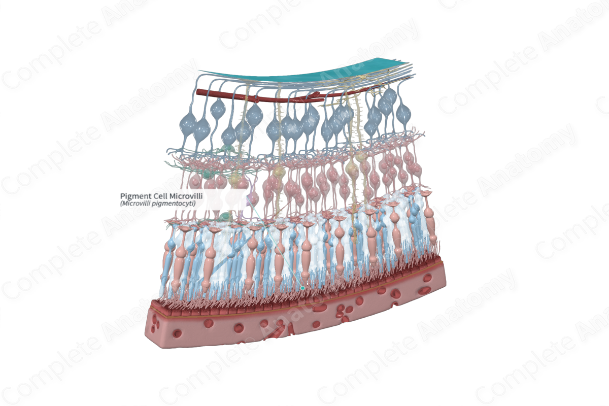
The pigmented layer of the retina is composed of a low cuboidal epithelium with long sheetlike apical microvilli. Measuring 5–7 µm in length, these microvilli interdigitate with photoreceptor cell outer segments (Standring, 2016).
The microvilli that surround the outer segments of cone photoreceptors are usually devoid of organelles while, in contrast, those surrounding rod photoreceptors are abundant in cellular organelles. Pigment cell microvilli house a core bundle of densely packed actin filaments. Myosin has been localized to the bases of apical processes (Bonilha et al., 2006).
Anatomical Relations
The apical microvilli extend from the inner or vitreal surface of the pigment epithelial cells. They extend to fill the spaces between the tips of the outer segments of photoreceptor cells, seeming to engulf them. Up to 30–40 microvilli may surround a single photoreceptor outer segment (Bonilha et al., 2006).
Function
The apical microvilli of the pigmented epithelium largely facilitate the pigmented layer’s phagocytic properties, as well as its ability to transport nutrients and removal of waste products into and out of the photoreceptor cells, respectively (Bonilha et al., 2006).
The apical microvilli extend across the potential space that is a remnant of the optic ventricle that becomes obliterated by the infolding that forms the optic cup. By interdigitating with and ensheathing photoreceptor outer segments to the microvilli contribute to the adhesion of the pigmented layer to the photoreceptors and the collective neural retina. This adhesion arises from frictional or electrostatic interactions (Bonilha et al., 2006; Marmor, 1993). Thus, the pigment epithelium has a role in preventing retinal detachment, and when such detachment occurs it does so along the potential space between the retinal pigment epithelium and the neural retina.
List of Clinical Correlates
- Retinal detachment
References
Bonilha, V. L., Rayborn, M. E., Bhattacharya, S. K., Gu, X., Crabb, J. S., Crabb, J. W. and Hollyfield, J. G. (2006) 'The retinal pigment epithelium apical microvilli and retinal function', Adv Exp Med Biol, 572, pp. 519-24.
Marmor, M. F. (1993) 'Chapter 8 Mechanisms of retinal adhesion', Progress in Retinal Research, 12, pp. 179-204.
Standring, S. (2016) Gray's Anatomy: The Anatomical Basis of Clinical Practice. Gray's Anatomy Series 41 edn.: Elsevier Limited.
