
Summertime beachgoers are all too familiar with the painful sting of a jellyfish. But how do the stinging cells of jellyfish, corals, and sea anemones actually work? New research from the Stowers Institute for Medical Research has unveiled a precise operational model for the stinging organelle—or nematocyst—of the starlet sea anemone, Nematostella vectensis. The study, headed by Ahmet Karabulut, a predoctoral researcher in the lab of Matt Gibson, PhD, involved the use of cutting-edge microscopic imaging techniques, along with the development of a biophysical model to enable a comprehensive understanding of a mechanism that has remained elusive for over a century.
The researchers suggest that insights from the work could help lead to new clinical developments, including the design of microscopic therapeutic delivery devices. “Understanding this complex stinging mechanism can have potential future applications for humans,” said Gibson. “This could lead to the development of new therapeutic or targeted delivery methods of medicines as well as the design of microscopic devices.” Gibson and colleagues reported on their study in Nature Communications, in a paper titled, “The architecture and operating mechanism of a cnidarian stinging organelle.”
The stinging organelles of jellyfish, sea anemones, and related cnidarian organisms are “remarkable cellular weapons,” which are used for both predation and defense, the authors wrote. Nematocysts consist of a pressurized capsule containing a coiled harpoon-like thread that delivers a cocktail of neurotoxins. “When triggered, the capsule discharges, ejecting its thread as a harpoon that penetrates targets, and rapidly elongates by turning inside out in a process called eversion,” the researchers explained. “At the cellular level, nematocyst discharge is among the fastest mechanical processes in nature, known to be completed within three milliseconds in Hydra nematocyst.” In fact, the initial phase of pressure-driven capsule explosion and subsequent thread ejection occurs in as fast as 700 nanoseconds.
Previous studies suggest that the high speed of nematocyst discharge is driven by the accumulation of osmotic pressure inside the capsule, and the elastically stretched capsule wall releasing energy by a powerful spring-like mechanism during discharge. “Upon triggering, but prior to discharge, the capsule approximately doubles in volume due to the rapid influx of water,” the authors said. “This causes the matrix to swell osmotically and stretches the capsule wall. This energy is subsequently utilized to eject the thread with high velocity, which impacts and penetrates target tissue.”
Although the nematocyst characteristics in different species of cnidarians do vary considerably with respect to capsule size and thread morphology, they all operate in a similar way, and feature an evertible tubule that is driven by explosive ejection.
The Stowers team’s model for stinging cell function provides crucial new insights into the detailed nature of the extraordinarily complex nematocyst architecture and firing mechanism. Karabulut and Gibson, in collaboration with scientists at the Stowers Institute Technology Centers, used advanced imaging, three-dimensional electron microscopy, and gene knockdown approaches to discover that the kinetic energy required for piercing and poisoning a target involves both osmotic pressure and elastic energy stored within multiple nematocyst sub-structures.
“We utilized fluorescence microscopy, advanced imaging techniques, and 3D electron microscopy combined with genetic perturbations to understand the structure and operating mechanism of nematocysts,” said Karabulut.
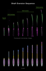
Using the state-of-the-art methods the researchers characterized the explosive discharge and biomechanical transformation of N. vectensis nematocysts during firing, into three distinct phases. The first phase is the initial, projectile-like discharge and target penetration of the densely coiled thread from the nematocyst capsule. This process is driven by a change in osmotic pressure from the sudden influx of water and elastic stretching of the capsule.
The second phase marks the discharge and elongation of the thread’s shaft substructure which is further propelled by the release of elastic energy through the eversion process—the mechanism where the shaft turns inside out—forming a triple helical structure to surround a fragile inner tubule decorated with barbs containing a cocktail of toxins. In the third phase, the tubule then begins its own eversion process to elongate into the soft tissue of the target, releasing neurotoxins along the way.

This entire stinging operation is completed within just a few thousandths of a second. “The earliest phase of the firing of the nematocyst is extremely fast and hard to capture in detail,” said Karabulut. As does sometimes happen in fundamental biological research, the initial discovery was an accident of curiosity. Karabulut incorporated fluorescent dye into a sea anemone to see what it looked like when the nematocyst-rich tentacles were triggered. After applying a combination of solutions to both activate nematocyst discharge and simultaneously preserve their delicate substructures in time and space, he found he had serendipitously captured multiple nematocysts at different stages of firing.
“Under the microscope, I saw a stunning snapshot of discharging threads on a tentacle. It was like a fireworks show. I realized nematocysts partially discharged their threads while the reagent I used simultaneously and instantaneously fixed the samples,” said Karabulut. “I was able to capture images that showed the geometric transformations of the thread during firing in a beautifully orchestrated process. After further examination, we were able to fully comprehend the geometric transformations of the nematocyst thread during its operation.”
“… this study demonstrates the operational capability of the nematocyst as a complex and self-assembling biological micromachine,” the authors concluded. “We propose that these ancient and sophisticated organelles represent an ideal model for biologically inspired microscale devices that could be utilized in diverse applications ranging from medical technology to materials science.”
